
GPCR activation monitoring: the GABY biosensors
How the biosensors work
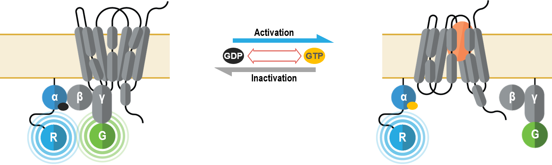
The Gα/γ-based G protein activation biosensors are used to monitor the activation of heterotrimeric G proteins at the plasma membrane upon receptor stimulation. Heterotrimeric G proteins are the canonical signaling partners of G protein-coupled receptors (GPCRs). Receptor activation triggers the exchange of a Gα-bound GDP for GTP, resulting in a conformational rearrangement of the heterotrimeric G protein that promotes dissociation of Gα and Gβ/γ subunits. In turn, GTP-bound Gα and free Gβ/γ subunits (β/γ remain associated) are then available to engage specific effectors. G proteins are grouped into families based on the signaling outcomes following activation of the Gα subunit. The Gs family (Gαs, Gαolf) bolsters the production of cAMP through direct activation of adenylyl cyclases (AC). Conversely, the Gi family (Gαi1, Gαi2, Gαi3, GαoA, GαoB and Gαz) reduces cAMP levels by inhibiting specific ACs. The Gq family (Gαq, Gα11, Gα14 and Gα15) activates Phospholipases Cβ (PLCβs) to produce the second messengers diacylglycerol (DAG) and inositol triphosphate (IP3), which subsequently promote the activation of Protein Kinases C (PKCs) and Ca2+ release from the endoplasmic reticulum, respectively. Finally, G12/13 family (Gα12 and Gα13) is known to control Rho-GEFs (Guanine nucleotide exchange factors) such as LARG, p115 and TRIO and thus influence processes linked to cytoskeletal remodeling (e.g., chemotaxis).
The bioSens-All® multimolecular GABY BRET sensors were designed to monitor the conformational changes that occur within the heterotrimeric complex upon Gα activation and effector interaction (1-3). Gα subunits are internally fused to Renilla luciferase (RLuc; R in figure below) and Gγ subunit is tagged at its N-terminus with green fluorescent protein (GFP; G in figure below). G protein activation by a receptor generally leads to a decrease in the BRET signal.
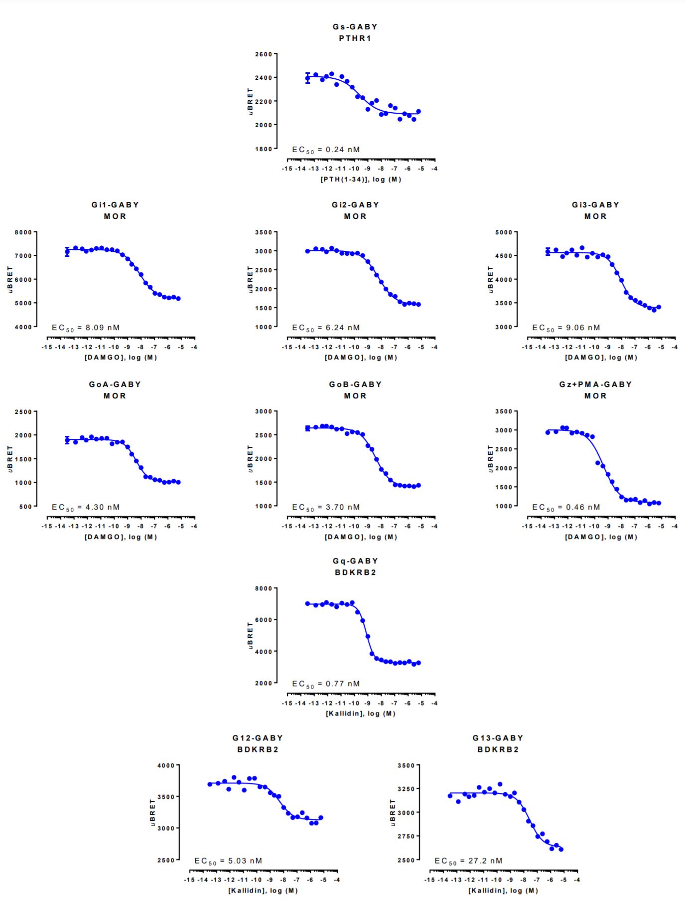
GABY biosensor data generated with the Parathyroid Hormone 1 receptor
HEK293 cells were transfected with a receptor coding plasmid (either human parathyroid hormone type 1 receptor (PTHR1), human mu opioid receptor (MOR) or human bradykinin receptor B2 (BDKRB2)) in addition to plasmids coding for the GABY biosensor. On the day of BRET, cells were rinsed with assay buffer, incubated with coelenterazine and increasing amounts of PTH(1-34), DAMGO or kallidin for 10 minutes and BRET subsequently measured.
References
1- Galés C, Rebois RV, Hogue M, Trieu P, Breit A, Hébert TE, Bouvier M. Real-time monitoring of receptor and G-protein interactions in living cells. 2005. Nat Methods. 2(3): 177-84. PMID 15782186.
2- Galés C, Van Durm JJ, Schaak S, Pontier S, Percherancier Y, Audet M, Paris H, Bouvier M. Probing the activation-promoted structural rearrangements in preassembled receptor-G protein complexes. 2006. Nat Struct Mol Biol. 13(9):778-86. PMID 16906158.
3- Busnelli M, Saulière A, Manning M, Bouvier M, Galés C, Chini B. Functional selective oxytocin-derived agonists discriminate between individual G protein family subtypes. 2012. J Biol Chem. 287(6): 3617-29. PMID 22069312.